
Translate this page into:
Non-invasive vital teeth whitening for enhanced facial esthetic: A case report
*Corresponding author: Pradyumna Misra, Department of Conservative Dentistry and Endodontics, Saraswati Dental College, Lucknow, Uttar Pradesh, India. drpmisra4lko@gmail.com
-
Received: ,
Accepted: ,
How to cite this article: Chauhan M, Singh K, Chauhan R, Shukla P, Misra P. Non-invasive vital teeth whitening for enhanced facial esthetic: A case report. Asian J Oral Health Allied Sci. 2023;13:8. doi: 10.25259/AJOHAS_24_2023
Abstract
For ages, a bright white smile symbolizes beauty, wellness, and vitality. The translucency and thickness of the enamel, hue of the underlying dentin, vitality, and color of the pulp all contribute to the pigmentation of teeth. Color changes in the crown can be due to physiologic or pathologic and endogenous or exogenous factors. Extrinsic or intrinsic stains are the most common causes of tooth discoloration. Discoloration, especially in the anterior teeth, seriously impairs esthetics and might make a patient feel less confident. In the treatment of discolored teeth, various invasive and non-invasive options, such as veneers and bleaching, are available, respectively. However, the urge for esthetic dentistry, particularly teeth whitening, has grown significantly in recent decades. As compared to invasive treatment options, non-invasive methods like bleaching (teeth whitening) are the most conservative choice for stained teeth and are preferred in situations where tooth structure is intact. Teeth whitening has developed into a well-liked and commonly requested dental procedure as a result of the public need for a brighter smile and enhanced looks. This paper aims to describe a case of vital, discolored anterior teeth due to fluorosis lightened using 35% hydrogen peroxide. A 19-year-old female patient reported to the department with discolored upper front teeth. After the procedure was completed, a pleasing esthetic result was achieved.
Keywords
Bleaching
Hydrogen peroxide
Tooth whitening
Vital

INTRODUCTION
Since esthetics have a significant impact on our culture, an imbalance in its harmony can have a detrimental effect on the quality of life by lowering self-esteem and disturbing social relationships. A patient’s confidence and dental health can both be greatly impacted by the esthetic improvement of their smile, and this can have a considerable positive impact on their overall well-being. Esthetics generally include form and color; discolored teeth may be viewed negatively in the concept of an ideal esthetic standard and may be challenging to treat.[1] Teeth discoloration factors can be categorized as intrinsic or extrinsic. Trauma can cause blood cells from the pulp to enter and stain the dentinal tubules, which, in turn, can change the color of healthy teeth, although it may not be severe enough to result in pulpal death. Trauma can also result in the partial occlusion of the pulp chamber or dentin deposition in it, known as calcific metamorphosis, which causes intrinsic discolorations.[2]
Dentists can restore discolored vital teeth using a variety of treatment options, which vary from highly invasive ones such as crowns and veneers and the placement of direct restorations to minimally invasive ones such as macroabrasion, microabrasion, and bleaching. Bleaching improves the appearance of the discolored teeth while preserving the sound tooth structure. It actually provides a safe, conservative approach for treating discolorations, satisfying the needs of both clinicians and patients, and is thus more widely accepted.[3]
Selection criteria for vital tooth bleaching
The concentration of the bleaching agent, length of use, the kind of tooth discoloration, color of the teeth, and the patient’s age are all significant elements that can affect the ultimate outcome after bleaching.[4]
According to reports, the following tooth discolorations have the best result for whitening:
Yellowing of teeth due to physical factors such as food, smoking, and aging
Staining due to fluorosis
Tooth darkening due to mild trauma
Tetracycline staining.
Many dentists include vital teeth bleaching into their esthetic bonding procedures. For patients who are unsatisfied with the unesthetic appearance of their discolored teeth, bleaching initially lightens the color of the teeth, making it easier to conceal tooth stains.
Before any bonding procedure, bleaching should be avoided for at least a week to prevent interference with adhesion in bonding and material setting.[5]
CASE REPORT
A 19-year-old female was referred to the Department of Conservative Dentistry and Endodontics with a chief complaint of unesthetic appearance due to discolored front teeth without any pain or symptoms. The patient had no systemic illnesses, and no drugs that stained teeth were being taken by her. Before any examination or treatment, a patient’s written informed permission was acquired, and a second written consent was also obtained for the presentation and documentation of the case. Subsequently, clinical and radiographic examinations were carried out. Diagnosis of dental fluorosis was made after taking the patient’s history and clinical examination [Figure 1].

- Pre-bleaching extraoral photograph.
Treatment procedure
First, a tooth vitality test was performed on maxillary anterior teeth using an electronic pulp vitality tester, and all teeth were found to be vital. Hence, a non-invasive bleaching procedure was decided for the patient. Photos were taken before starting the procedure [Figure 1]. The patient initially had a shade 3L-1.5, which was verified by a VITA ToothGuide 3D Master (Vita, Germany).
Before beginning the bleaching procedure, oral prophylaxis was performed. About 35% hydrogen peroxide (Pola Office Plus, SDI, Australia) was the chosen treatment material for the patient [Table 1]. This product predominantly contains hydrogen peroxide and silicon dioxide powder that has proven oxidizing action, and also has potassium nitrate that acts as a desensitizer. The treatment procedure started with isolating the field, followed by the application of separating media (Vaseline) on soft tissues. After this, a gingival barrier was applied and light cured for 20 s [Figure 2]. Pola Office Plus syringe was fitted with a tightly clamped tip, and the plunger was gently drawn back to release the pressure. According to the manufacturer’s instructions, the syringe contents were gently released into the pot and blended as soon as possible, using a brush applicator until the gel was uniform. Post this, the gel was applied to all surfaces of the teeth for eight minutes, followed by light activation using Coltolux LED (Coltene Whaledent, Germany) [Figure 3].
| Material | Composition |
|---|---|
| 1. Pola office plus (pH=7) | Pola office liquid
Pola office powder
Gingival barrier
|
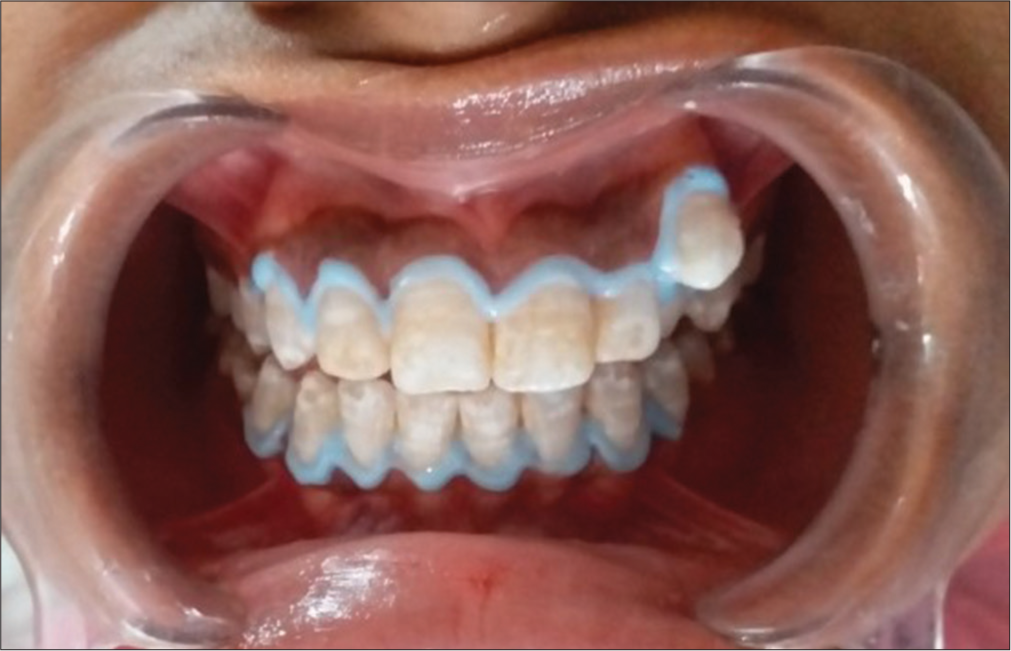
- Application of gingival barrier.

- Application of gel with brush applicator.
A surgical aspirator tip was used for suction. Two cycles were performed in one session. The bleaching agent was removed using an air-water syringe and suction [Figure 4].

- Post bleaching extraoral photograph.
The patient was recalled after one week to assess the outcomes. A visible shade change was seen when examined with standard visual examination protocol, and verified with VITA Tooth guide 3D Master, and the post-operative shade changed to 3M-1. Once the necessary shade enhancement was achieved, final polishing was done to achieve a smooth surface.
DISCUSSION
Nearly all instances of tooth discoloration, including those caused by pulp tissue disintegration, internal hemorrhage, damage to the primary dentition, drug usage, restorative materials, and systemic disorders, including fluorosis and jaundice, require bleaching. Pregnant women, newborns, children under the age of 10, people with open dentinal tubules, and those who can’t stop smoking during treatment are not advised to undergo the bleaching operation. Regardless of the bleaching technique or product used, diffusion of peroxide into the organic matter of tooth structure relies on the diffusion coefficient, extent of application, and concentration of the active bleaching agent. By heating up the bleaching agent, photocatalytic interactions increase the breakdown of hydrogen peroxide and thus boost the association between reactive oxygen and pigmented molecules, leading to intensified bleaching efficiency.[6]
Before starting treatment, there are some rules that must be followed to prevent discomfort caused by the procedure. This includes refraining from eating or drinking anything that is excessively hot or cold, anything with a low Potential of Hydrogen (pH), quitting smoking, and engaging in any activity that can make teeth more sensitive.[7] To perform dental office whitening with improved safety and effectiveness, a new generation of bleaching products that employ low hydrogen peroxide concentrations (3.5% and 15%) was introduced into the market.
The bleaching procedure is one of several esthetic treatment modalities available in dentistry today. There are various brands and concentrations of bleaching agents available in the market. In this case, Pola Office Plus bleaching was used and the results were promising. Along with 35% hydrogen peroxide, the Pola Office Plus contains potassium nitrate that reduces the patient’s post-bleaching sensitivity.
Before bleaching, a proper clinical evaluation and history are essential to determine the etiological factor responsible for tooth discoloration and the degree of discoloration. Non-vital teeth and traumatic injuries to teeth also cause tooth discoloration; therefore, vitality testing should be performed before the bleaching procedure to avoid incorrect diagnosis and treatment.
Increased bleaching agent concentration is harmful to pulp tissue. Different in vitro studies have shown that bleaching agent penetration into the pulp chamber occurs after 60 min of bleaching agent exposure to the tooth surface. According to Hanks et al.,[8] bleaching agent penetration into the pulp chamber depends on the bleaching agent’s initial concentration and the length of time it was applied to the tooth surface; he also came to the conclusion that the process required around 15 min.
The molecule of peroxide has a very small molecular size and weight and has the ability to denature the protein present in dentin, which allows it to move easily through dentinal tubules and reach the pulp chamber. Two other elements that aid in bleaching agent diffusion into the pulp chamber are the positive pressure inside the chamber and the osmotic pressure of the bleaching agent.[9] However, in vivo studies yield the opposite result as in vitro studies. Cohen and Robertson’s in vivo studies revealed either no or very little pulpal inflammation when exposed to 35% hydrogen peroxide.[10,11] Peroxidase and catalase enzymes found in pulp provide protection against bleaching agents by degrading hydrogen peroxide molecules.
CONCLUSION
Vital tooth whitening is a non-invasive treatment option that may significantly enhance the appearance of teeth. Clinical findings support the use of professional tooth whitening as a practical and attractive therapy for stained dentition. It is a crucial component of a cosmetic dentistry treatment plan due to its low risk and lack of invasiveness. The general population has become more interested in in-office bleaching. Nowadays, a lot of patients are aware that in-office bleaching is a service that many dentists provide, as it is a great way to attain fast and detectable changes in tooth color.
Ethical approval
The Institutional Review Board approval is not required.
Declaration of patient consent
The authors certify that they have obtained all appropriate patient consent.
Conflicts of interest
Dr. Raju Chauhan is on the Editorial Board of the Journal.
Use of artificial intelligence (AI)-assisted technology for manuscript preparation
The authors confirm that there was no use of artificial intelligence (AI)-assisted technology for assisting in the writing or editing of the manuscript and no images were manipulated using AI.
Financial support and sponsorship
Nil.
References
- Dental bleaching and new possibilities-literature review. Health Sci J. 2018;12:600.
- [CrossRef] [Google Scholar]
- Nightguard vital bleaching: Current concepts and research. J Am Dent Assoc. 1997;128:19-25.
- [CrossRef] [PubMed] [Google Scholar]
- Time duration for dissipation of bleaching effects before enamel bonding. J Dent Res. 1992;71:179-85.
- [Google Scholar]
- Effects of the hydroxyl radical and hydrogen peroxide on tooth bleaching. J Endod. 2004;30:45-50.
- [CrossRef] [PubMed] [Google Scholar]
- Human pulp responses to in-office tooth bleaching. Oral Surg Oral Med Oral Pathol Oral Radiol Endod. 2010;109:59-64.
- [CrossRef] [PubMed] [Google Scholar]
- Cytotoxicity and dentin permeability of carbamide peroxide and hydrogen peroxide vital bleaching materials, in vitro. J Dent Res. 1993;72:931-8.
- [CrossRef] [PubMed] [Google Scholar]
- Penetration of 38% hydrogen peroxide into the pulp chamber in bovine and human teeth submitted to office bleach technique. J Endod. 2007;33:1074-7.
- [CrossRef] [PubMed] [Google Scholar]
- Human pulpal response to bleaching procedures on vital teeth. J Endod. 1979;5:134-8.
- [CrossRef] [PubMed] [Google Scholar]
- Pulpal response to vital bleaching procedures. J Endod. 1980;6:645-9.
- [CrossRef] [PubMed] [Google Scholar]


