
Translate this page into:
The necessity of radiographic investigation before dental extraction from a student’s perspective
-
Received: ,
Accepted: ,
How to cite this article: Jiboon AT, Alsheakh AJ, Alhamdani F, Hussein M, Waleed Z, Saad T. The necessity of radiographic investigation before dental extraction from a student’s perspective. Asian J Oral Health Allied Sci 2022;12:11.
Abstract
Objectives:
The objectives of this study were to determine students’ points of view regarding the necessity of dental radiography for examination.
Material and Methods:
A Google form questionnaire formulated by A. J. and F. A. was circulated among dental students, through dental students’ channels on Telegram, Instagram, and Facebook for 26 days (from December 10, 2021, to January 4, 2022). The questionnaire contains six questions on what is thought to be relevant to the study’s aim. The questions were based on 16 years of educational experience in clinical training in the oral surgery clinic. Students from Baghdad, Basra, Anbar, and Babylon provinces participated in this study. Contributions were from governmental and private dental schools. The questionnaire constituted six items investigating aspects of radiographic investigations concerning dental extraction, as viewed by dental students.
Results:
Two hundred and fifty-four students and new graduates answered the questionnaire. The overwhelming majority (67.3%) of participants stated that a radiograph is needed for all teeth before dental extraction. Seventy-five out of 180 participants think that an X-ray is required to evaluate the relationship with vital structure. Out of 254, 163 participants preferred a periapical radiograph before dental extraction followed by OPG. The change in the tendency toward X-ray type during the academic year has been statistically confirmed (P = 0.002).
Conclusion:
Students and new graduates seem to favor taking X-ray before dental extraction only if needed. It seems to suggest that with the progress of the study, participants are more inclined to choose problem-based X-ray examinations. The screening purpose of X-ray investigation seems to be overlooked with the progress of study and practice.
Keywords
Dental extraction
Dental radiography
Dental students
Periapical radiography
OPG
CBCT

INTRODUCTION
A dental extraction is the removal of teeth from the dental alveolus (socket) in the alveolar bone for various pathological reasons. A dental extraction is the most common oral surgical procedure in dental practice.[1] Mastering dental extraction is an essential prerequisite for a qualified dentist. Proper dental extraction mandates comprehensive condition assessment to perform the procedure efficiently with minimum complications[2] and performing the procedure with the least possible trauma to the surrounding tissue.[3]
One of the most important investigations before dental extraction is dental radiography.[4] It provides information about the roots and the surrounding tissues.[5] It, also, provides detailed information about the presence of ankylosis with replacement resorption or hypercementosis, pathology in the surrounding bone, relationship with the adjacent roots, and vital structure.[6]
According to the Iraqi Dental School, there is no strict role regarding the need for dental radiography before dental extraction. In most cases, the decision to take an X-ray before extraction is based on the senior judgment on whether there is a need to evaluate the tooth or the jaw bones. This study aims to assess dental students’ perspective toward radiographic examination before conventional dental extraction.
MATERIAL AND METHODS
A Google form questionnaire on an X-ray before dental extraction was formulated by A. J. and F. A. and circulated among dental students’ channels on Telegram, Instagram, and Facebook for 26 days (from December 10, 2021, to January 4, 2022). The questionnaire contains six questions on what is thought to be relevant to the study’s aim. The questions were based on 16 years of educational experience in clinical training in the oral surgery clinic. Participants were from different Iraqi governmental and private dental schools in Baghdad, Basra, Anbar, and Babylon governorates. This questionnaire was directed to the 4th- and 5th-year students and newly graduated dentists [Appendix 1]. The newly graduated dentists are the past year graduates who have not yet started their postgraduate training work as house officers. Only responses from students who had finished their first semester working with patients in the oral surgery department clinic were included in the study. Responses from students from other academic years were excluded from the study.
The questionnaire constituted of six items investigating various aspects of radiographic investigation about dental extraction, as viewed by the dental student. The questionnaire focuses on the student’s opinion of the necessity of radiographic imaging.
Both descriptive and inferential statistics were used for statistical analysis. Chi-square test was used to identify the relationship between nominal, and ordinal variables. Spearman Correlation Test was used to examine the relationship between ordinal variables. P < 0.05 was considered the limit for the statistical significance. IBM® SPSS, Ver 25 was used for statistical analysis.
RESULTS
Two hundred and fifty-four students and new graduates answered the questionnaire. Study descriptive statistics for the number and percentages in terms of gender, the academic year, the necessity of X-ray, teeth for which X-ray is required, and the type of X-ray investigation are shown in [Table 1].
| Variable | Number | Percentage |
|---|---|---|
| Gender | ||
| Males | 81 | 31.9 |
| Females | 173 | 68.1 |
| Academic year | ||
| 4th-year students | 68 | 26.8 |
| 5th-year students | 153 | 60.2 |
| Newly graduates | 33 | 13 |
| The need for an X-ray before dental extraction | ||
| Not required | 6 | 2.4 |
| On need | 171 | 67.3 |
| All cases | 77 | 30.3 |
| Teeth where an X-ray is required for extraction | ||
| Wisdom teeth | 44 | 17.3 |
| X-ray for molars | 21 | 8.3 |
| X-ray for premolars and molars | 58 | 22.8 |
| X-ray for all teeth | 131 | 51.6 |
| Type of X-ray required before dental extraction | ||
| Periapical | 163 | 64.2 |
| OPG | 72 | 28.3 |
| CBCT | 19 | 7.5 |
As expected, the female-to-male ratio was 2/1. Out of 254 participants, about two-thirds were 5th-year students. About a quadrant of the respondents were 4th-year students. The remaining number belongs to the new graduate dentists.
[Table 1] clarifies students’ opinions regarding the need for an X-ray before extraction. The overwhelming majority of participants stated that a radiograph is needed for all teeth before dental extraction, followed by participants who think that radiography is needed in certain cases. Very few respondents see that it is not necessary to take an X-ray before dental extraction. Statistical analysis did not show a significant relationship between the need for X-ray and participant gender (P = 0.232) or participant academic year (P = 0.566).
More than half of the respondents indicate the need to take an X-ray of all teeth before dental extraction; when the other half divided between a specific type of teeth, premolars and molars got more than one-third, and wisdom teeth were voted by less than third of participants, whereas the least number of votes considered molar teeth for X-ray investigation. There is no statistically significant relationship between the academic year and the tooth for which an X-ray was requested (P = 0.937). Furthermore, no significant relationship between the type of requested X-ray and the condition the X-ray is requested for.
Approximately two-thirds of the participants preferred a periapical radiograph before dental extraction, followed by OPG, which is preferred by about a third of the participants. A minority of the participants chose CBCT to be the radiographic tool for investigation before dental extraction.
As shown in [Figure 1], 75 out of 180 participants think that an X-ray is required to evaluate the relationship with vital structure. Root configuration was considered by 44 participants to be the reason for requesting radiographic examinations. This was followed by the presence of periapical pathology and the presence of unexpected pathology (30, and 24 participants, respectively). Only seven participants prefer to take an X-ray for medicolegal consideration. There was no statistically significant relationship between the condition, for which an X-ray was requested and the type of radiographic investigation (P = 0.2).
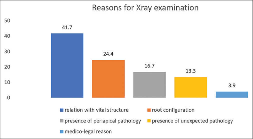
- Percentage of responses regarding the condition for which X-ray is required.
What [Figure 2] clearly shows the tendency toward periapical X-ray among students as they progress in their clinical training. Panoramic view almost shares the same percentage in preference with periapical radiography for 4th-year students, whereas it is less widely considered for 5th-year students and newly graduated dentists. CBCT, on the other hand, is the least popular choice by the participants. The choice for CBCT with the advent of clinical training until it is no longer chosen by newly graduated dentists before dental extraction. The change in the tendency toward X-ray type during the academic year has been statistically confirmed (P = 0.002).

- The choice of X-ray type by academic year.
DISCUSSION
This study seeks to evaluate radiographic investigation before routine dental extraction procedures by dental trainees during their past 2 years of academic training and new graduates who are about to start their professional careers. To the best of our knowledge, this is the first study in Iraq in this field. It is worth considering; however, that students’ evaluation reflects a certain extent the educational institute’s view regarding any given training material. Needless to mention that clinical dental education has been significantly affected by COVID19 lockdown in the past 2 years.[7]
As the study shows, females represent the majority of the participants. This has been a consistent finding in more than one Iraqi study.[7] Passion is one of the factors. However, the choice might be influenced by the parents’ preference. Dentistry is less demanding compared to medical schools, with minimum patient risk, and shorter in terms of study years compared to medical school. It is also considered a more comfortable career choice.
Performing, interpreting dental radiography, and making decisions based on their interpretation are the responsibilities of the dentist. It is part of the requirements of general dental practice.[8] Radiographic examination, by default, is requested on need after history taking and clinical examination. Clinical examination governs the type of required radiograph and the area to be examined to confirm or exclude one or more of the differential diagnostic options. In another word, the radiographic investigation is dictated by the clinical decision. Radiographs by themselves are suggestive, not diagnostic tools.
In Iraqi dental schools, oral surgery training usually focuses on dental extraction procedures, mostly straightforward dental extraction procedures. Radiographic investigation in most oral surgery departments in Iraqi dental schools is usually requested for wisdom teeth when a difficulty or complication is faced during the surgical procedure. This may explain the high percentage of preferred pre-extraction radiography for certain cases. Participants who preferred not to take an X-ray before dental extraction might be influenced by their training, in which an X-ray was not requested for the teeth that they extracted.
According to the ADA regulations, there are no standard requirements for diagnostic radiographic investigations. A dentist’s judgment based on the patient’s need seems the base for ordering an X-ray. The radiographic investigation is considered as a resource rather than a standard care requirement.[9] Similarly, the Faculty of General Dental Practice in the UK states that there is no compelling evidence to support the need for routine radiography before dental extractions in adults. However, it would be sensible to request a radiograph in certain situations.[10]
Fear of procedure-related complications seems to influence the need for an X-ray. Vital structures such as maxillary sinus and inferior alveolar nerve[11,12] might interfere with the surgical decision. This might, also, justify the choice of responses regarding pre-extraction X-ray for molar and premolar teeth.[13-15] The other reason for requesting an X-ray in multi-rooted premolar and molar teeth could be related to their roof configuration. Variations in root shape, angulation, and the possible need for root sectioning make radiographic examination understandable.
Tooth-related periapical or unexpected pathologies did not seem to raise the same concern for dental radiography. The request for the X-ray to identify the presence of a periapical lesion, however, might be related to the possible influence of periapical inflammatory lesions on the effect of local anesthesia, which might result in a painful dental extraction procedure.
The presence of unexpected pathology such as cysts/tumours, impacted teeth, or retained roots. This type of information is very important before starting any surgical procedure, because, first, it will enhance the decision whether to extract the tooth or not, in addition, it might influence the surgical procedure, we might need to alter the surgical procedure according to the radiographic findings.[16]
The preference of participants for periapical X-ray could be related to their availability. It is simple to use and does not require professional expertise and can provide the needed information for the tooth in question and the surrounding structures. In addition, the use of modern equipment combined with good technique and sensitive detectors can minimize the risk of radiation.[17]
Nevertheless, a periapical radiograph is not suitable for screening purposes, when multiple extractions are indicated. This is why some of the participants chose OPG as an investigation tool before dental extraction. It is suitable for multiple teeth extractions, which is not uncommon for oral surgery department patients. Panoramic radiograph reduces the need for multiple radiographs and X-ray exposure. Besides, it is useful for limited that mouth opening or the bone level is not revealed enough. OPG reflects the complete arch image containing the teeth, bone, and related pathologies.[18-20] It is, also, available in all dental teaching facilities.
The decrease in interest in OPG with the progress in the academic study might reflect that students are becoming more interested in the tooth in question rather than screening the purposes. The aforementioned clinical training environment appears to play a role in encouraging problem-focused radiographic investigation. The choice of X-ray type might also indicate that practical thinking is more evident. Despite the presence of CBCT in the majority of dental schools, CBCT is not requested for what is deemed straightforward dental extraction, because it is expensive and unavailable in most dental clinics with high radiation doses. Furthermore, it is difficult for undergraduate students or newly graduated dentists to interpret three-dimensional images.
In terms of academic practice in Iraq, it does not seem that there is no generally implemented protocol regarding the use of dental radiology before dental extraction. Besides, the type of radiographic technique is not specified. Some references refer to dental radiographs.[21] Others prefer a panoramic view,[22] or even CBCT.[23] Dental radiographs in Iraqi academic institutes are not requested for all teeth indicated for extraction.
A minority of participants chose medicolegal reasons for pre-extraction radiography. In terms of medicolegal obligation, the dentist should inform the patient about the available treatment option in the light of the best diagnostic evidence, allowing the patient to be involved in the treatment decision,[[24] and protects the dentist from any claim of negligence.[25] Failure to take an X-ray before dental extraction might put the dentist at risk of negligence claims.
This questionnaire is investigatory-based rather than practice based. Doing comparison between the need for radiographic investigation and the difficulty of extraction was out of the scope of the study.Besides, in oral surgery clinic in almost all oral surgery clinic in Iraqi dental schools, radiographs are not a routine practice and they are requested on limited situations based on the tutor’s opinion.
To summarize, students and new graduates seem to favor taking X-ray before dental extraction only if needed. It seems to suggest that with the progress of the study, participants are more inclined to choose problem-based X-ray examinations. The screening purpose of X-ray investigation seems to be overlooked with the progress of the study.
Declaration of patient consent
Patient’s consent not required as there are no patients in this study.
Financial support and sponsorship
Nil.
Conflicts of interest
There are no conflicts of interest.
References
- Influence of trans-operative complications on socket healing following dental extractions. J Contemp Dent Pract. 2007;8:52-9.
- [CrossRef] [PubMed] [Google Scholar]
- Dentoalveolar Surgery. In: An Issue of Oral and Maxillofacial Surgery Clinics of North America. E-Book. Vol 32. New York: Elsevier Health Sciences; 2020.
- [CrossRef] [Google Scholar]
- Are systemic antibiotics necessary in the prevention of wound healing complications after intra-alveolar dental extraction? Int J Oral Maxillofac Surg. 2016;45:1658-64.
- [CrossRef] [PubMed] [Google Scholar]
- Basic Guide to Oral and Maxillofacial Surgery (1st ed). Chennai, India: John Wiley and Sons, Ltd; 2017.
- [Google Scholar]
- Principles of routine exodontia In: Tucker M, Ellis E, eds. Contemporary Oral and Maxillary Surgery (7th ed). Philadelphia, (PA): Elsevier; 2019.
- [Google Scholar]
- Dentoalveolar surgery In: Pogrel KE, Kahnberg, L Andersson, eds. Essentials of Oral and Maxillofacial Surgery (1st ed). Chichester, West Sussex: John Wiley & Sons, Ltd, The Atrium, Southern Gate; 2014.
- [Google Scholar]
- Oral surgery learning outcome during Covid-19 lockdown a student-based evaluation. J Res Med Dent Sci. 2021;9:307-11.
- [Google Scholar]
- Contemporary medico-legal dental radiology. Aust Dent J. 2012;57:9-15.
- [CrossRef] [PubMed] [Google Scholar]
- X-Rays/Radiographs - American Dental Association. Department of Scientific Information, E.S.T.R., ADA Science and Research Institute, LLC.
- [Google Scholar]
- Selection Criteria for Dental Radiography England: Faculty of General Dental Practice UK; 2018.
- [Google Scholar]
- Clinical assessment of the relationship between the third molar and the inferior alveolar canal using panoramic images and computed tomography. J Oral Maxillofac Surg. 2008;66:2308-13.
- [CrossRef] [PubMed] [Google Scholar]
- Evaluating the risk of post-extraction inferior alveolar nerve injury through the relative position of the lower third molar root and inferior alveolar canal. Int J Oral Maxillofac Surg. 2019;48:1577-83.
- [CrossRef] [PubMed] [Google Scholar]
- Evaluation of the relationship between maxillary posterior teeth and the maxillary sinus floor using cone-beam computed tomography. BMC Oral Health. 2018;18:164.
- [CrossRef] [PubMed] [Google Scholar]
- Proximity of maxillary posterior teeth roots to maxillary sinus and adjacent structures using denta scan®. Indian J Dent. 2016;7:126-30.
- [CrossRef] [PubMed] [Google Scholar]
- Correlation between maxillary sinus floor topography and related root position of posterior teeth using panoramic and cross-sectional computed tomography imaging. Oral Surg Oral Med Oral Pathol Oral Radiol Endod. 2006;102:375-81.
- [CrossRef] [PubMed] [Google Scholar]
- Dental Radiography-E-book: Principles and Techniques New York: Elsevier Health Sciences; 2021.
- [Google Scholar]
- Is it true that the radiation dose to which patients are exposed has decreased with modern radiographic films? Dentomaxillofac Radiol. 2009;38:92-7.
- [CrossRef] [PubMed] [Google Scholar]
- Assessment of panoramic radiography as a national oral examination tool: Review of the literature. Imaging Sci Dent. 2011;41:1-6.
- [CrossRef] [PubMed] [Google Scholar]
- Diagnostic accuracy of panoramic radiography and ultrasonography in detecting periapical lesions using periapical radiography as a gold standard. Dentomaxillofac Radiol. 2020;49:20190290.
- [CrossRef] [PubMed] [Google Scholar]
- Basic and Complex Exodontia and Surgical Management of Impacted Teeth In: Fonseca Oral and Maxillofacial Surgery (3rd ed). Riverport Lane St. Louis, Missouri: Elsevier; 2018. p. :3251.
- [Google Scholar]
- Routine Extraction of Teeth. In: Kademani D, Tiwana PS, eds. Atlas of Oral and Maxillofacial Surgery. Riverport Lane St. Louis, Missouri: Elsevier; 2016. p. :3251.
- [Google Scholar]
- Surgical Extractions In: Mehra P, D'Innocenzo R, eds. Manual of Oral Surgery for the General Dentist (2nd ed). New Jersey: John Wiley and Sons Inc; 2016.
- [Google Scholar]
- Principles of Dentoalveolar Extractions In: The Atrium. Southern Gate: John Wiley & Sons Ltd; 2021. p. :PO19, 8SQ.
- [CrossRef] [Google Scholar]
- Professionalism and Ethics In: Frost DE, Le B, Powers MP, eds. Fonseca, Oral and Maxillofacial Surgery Vol 1. (3rd ed). Riverport Lane St, Louis: Elsvier; 2018. p. :3251.
- [Google Scholar]
- Legal modalities in dental patient management and professional misconduct. SRM J Res Dent Res. 2014;5:91.
- [CrossRef] [Google Scholar]
APPENDIX 1
Academic year Fourth Year Fifth Year Newly graduate Gender Male Female Is radiography required before extraction during undergraduate study? Yes No On need If your answer is (on need), what is the condition you prefer to take X-ray for:
Root configuration Presence of periapical lesion Presence of unexpected pathology Relationship with vital structure (maxillary sinus, inferior alveolar nerve) For medicolegal purpose I didn’t vote (on need) In your opinion for what teeth we need to take X-ray for? All teeth Premolars and molars Molars only Wisdom teeth only What are the type of X-ray you think is most appropriate before dental extraction? Periapical X-ray OPG CBCT


