A simplified approach for management of flabby ridge
*Corresponding author: Saumya Nagar, Departments of Prosthodontics and Crown & Bridge, Saraswati Dental College and Hospital, Lucknow, Uttar Pradesh, India. saumya2nagar@gmail.com
-
Received: ,
Accepted: ,
How to cite this article: Nagar S, Yadav R, Gaur A, Chopra D. A simplified approach for management of flabby ridge. Asian J Oral Health Allied Sci. 2025;15:4. doi: 10.25259/AJOHAS_17_2024
Abstract
For individuals with “flabby” or “fibrous” alveolar ridges, creating stable and retentive dental prostheses presents substantial challenges. Problems specifically occur when forces cause the movable tissues that support the denture to become deformed during the impression-taking process. A prosthodontist faces challenges when rehabilitating a patient who is entirely edentulous since flabby tissue negatively impacts the stability, support, and retention of complete dentures. To assist in resolving the issues that arise in these situations, a number of imprint approaches have been put forth. The simplest and most dependable maxillary anterior flabby ridge care has been demonstrated in this case.
Keywords
Combination syndrome
Flabby tissue
Impression
Kellys syndrome
INTRODUTION
A superficial region of movable soft tissue influencing the maxillary or mandibular alveolar ridges was referred to as a “flabby” or “fibrous” ridge. It was a common discovery, especially in long-term denture wearers, and can arise when hyperplastic soft tissue replaces the alveolar bone. Trauma from denture bases is said to be the cause of these ridges.[1]
It was more frequently discovered in the anterior area in the edentulous patient. Kelly originally identified “combination syndrome” in 1972 based on observations made during a 3-year follow-up period with six patients. Every patient had a removable partial denture that extended distally and a complete maxillary denture that was opposed by mandibular teeth.[2]
He noted the growth of the tuberosities, the resorption of alveolar bone in the front maxilla, and the resorption of bone beneath the bases of the mandibular dentures. Histologically, hyperplastic mucosal tissue, loosely distributed fibrous connective tissue, and robust collagenous connective tissue make up flabby ridges. There is a significant quantity of visible metaplastic cartilage and/or bone in the soft tissue.[3]
CASE REPORT
A 58-year-old male patient reported to the Department of Prosthodontics, Saraswati Dental College and Hospital, Lucknow, UP, India, with a chief complaint of missing teeth in the upper and lower jaw. An intraoral examination revealed edentulous maxillary and mandibular arches, high, well-formed, regular, and smooth maxillary and mandibular arches, and fibrous tissue in the anterior region of the maxillary arch [Figure 1a and b]. The patient previously had worn a denture for six years.
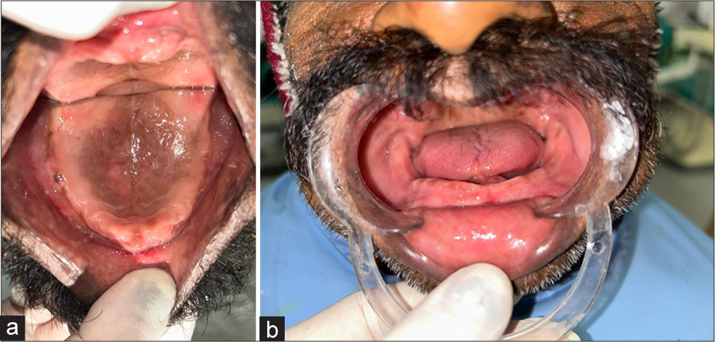
- (a) Maxillary ridge and (b) mandibular ridge.
The patient expressed apprehension regarding surgical procedures, such as the removal of loose tissue or treatment of implants, after evaluating the choices for their treatment plan. The patient’s maxillary and mandibular complete dentures were to be fabricated as part of the selected treatment plan.[4]
Selection of impression
To minimize the distortion of the flabby tissue, the admix technique was used to make the primary imprint of the mandibular denture bearing and the maxillary denture bearing area [Figures 2a and b]. Impressions were poured with dental plaster. The flabby tissue area was marked with an indelible pencil in the maxillary arch, and later, the area was measured with a thread.
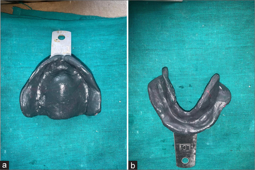
- (a) Maxillary primary impression and (b) mandibular primary impression.
Fabrication of wax spacer
The incisive papilla and the midpalatine raphe areas received relief wax, the flabby tissue area received two sheets of wax spacer, and the remaining areas received a single layer of wax spacer [Figure 3].
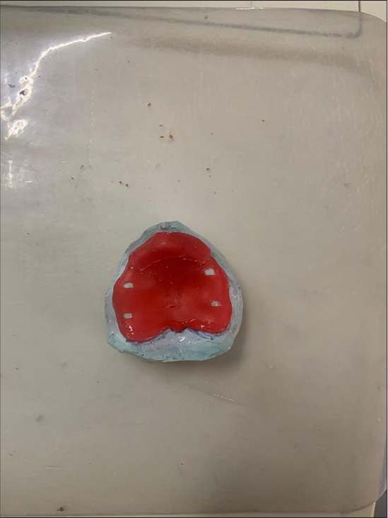
- Wax spacer for maxilla.
Recording of flabby tissue
Using autopolymerizing acrylic resin, a tray was made to which a little handle was attached. Green Stick compound (Pinnacle, DPI, India) was used for border molding [Figure 4]. After the spacer was removed from the undisplaced area, zinc oxide eugenol was used to make an impression [Figure 5a and b].[5]
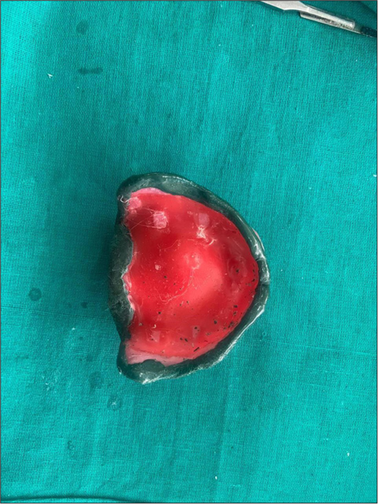
- Maxillary border molding.
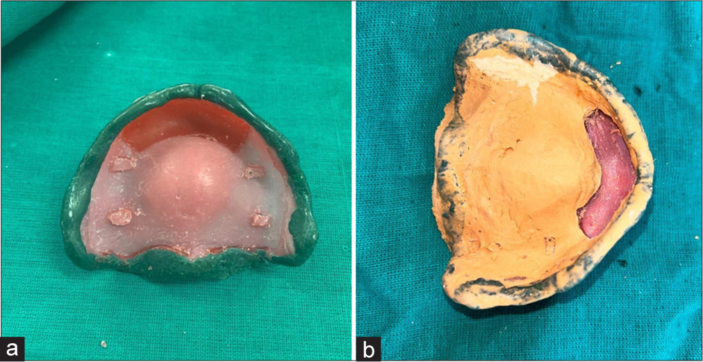
- (a) After removal of spacer and (b) ZOE impression of non-flabby tissue. ZOE: Zince oxide eugenol.
To prevent the flabby tissues from being squeezed, two holes were made on the rugae area of the flabby tissue using a straight fissure bur that was 2 mm wide [Figure 6] following the tray adhesive’s application (Universal tray adhesive, Zhermack, Italy). After loading the medium body into a syringe, material was injected through the holes created on the rugae. Extra material flowed from the other hole [Figure 7a and b]. A dental stone was used to pour in the impression.
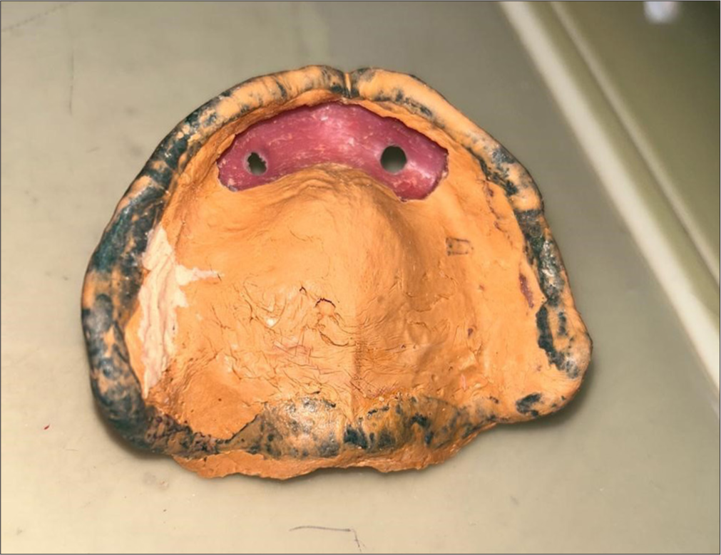
- A 2 mm wide hole made in the flabby tissue area.
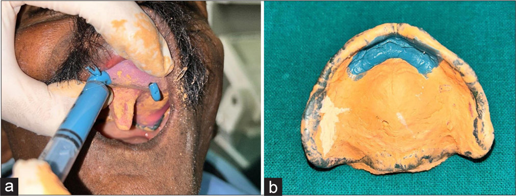
- (a) Medium body material injected through syringe and (b) maxillary secondary impression with medium body in flabby area.
A permanent base made of heat-cured acrylic resin was constructed. The cast was articulated in a semi-adjustable articulator (Hanau wide view, US) after the face-bow transfer. The goal of the teeth arrangement was balanced occlusion [Figure 8]. After processing, the denture was inserted [Figure 9]. The maxillary denture’s stability and retention were assessed clinically. The patient was said to be satisfied with the complete denture’s function, appearance, and retention at the subsequent appointments.[6,7]
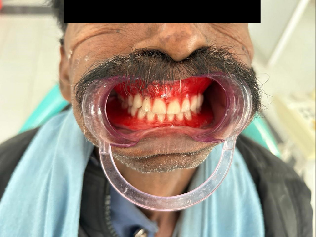
- Try – in.
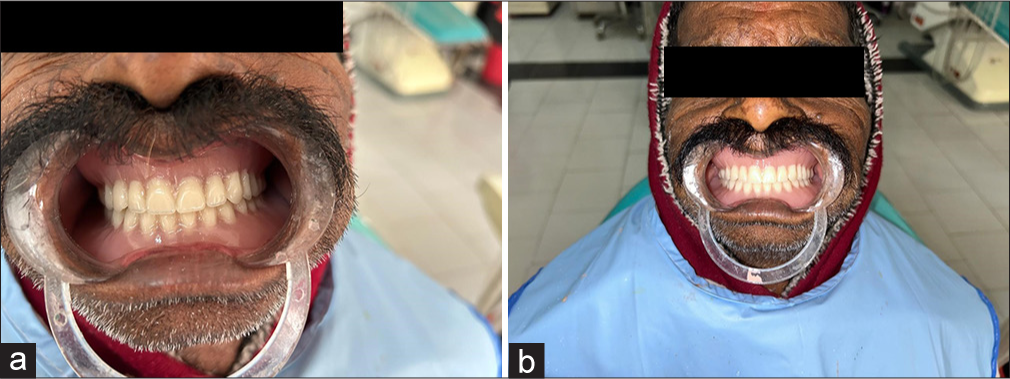
- (a and b) Denture insertion.
DISCUSSION
A good prosthodontic therapy, either by itself or in an interdisciplinary combination with surgery, can effectively correct flabby ridges. Provided there is sufficient bone height, it is possible to surgically remove flabby tissue.[8] However, it also causes a shallow sulcus depth, necessitating a minor surgical procedure called vestibuloplasty. Ridge augmentation is a possible remedy for this; however, it results in graft rejection or resorption once more. It has been suggested that sclerosing drugs like sodium morrhuate be injected into these kinds of flabby tissues, making them firm. Nevertheless, adverse responses, patient pain, and a decrease in hardness are among the documented consequences associated with these sclerosing drugs. Dentures made using conventional impression procedures or usually using window technique to capture such flabby tissues frequently become unstable and require additional time to create an accurate window for impression. This technique minimizes the distortion/displacement of hypermobile tissues by imprinting flabby areas and lowering hydraulic pressure. When making a secondary impression, this is a reliable method for controlled application of impression material.[9,10]
Clinical significance
This technique does not require additional clinical steps or more time than conventional impression techniques. For flabby ridge impressions, medium body impression materials have shown promising results in minimal tissue displacement, improved denture-related outcomes, and increased patient satisfaction.
CONCLUSION
It can be difficult to manage a patient with a flabby maxillary ridge, and good denture management requires careful consideration of the impact of both the impression surface and the occlusal surface. Using standard muco-compressive impression procedures is likely to produce a denture that is unstable and unretentive. Mucostatic impression procedures might not optimize the utilization of available tissue support, and there might be concerns with the denture base’s mobility in relation to the support tissues. Several of these restrictions should be addressed by the application of selective pressure impression techniques.
Ethical approval
The Institutional Review Board approval is not required.
Declaration of patient consent
The authors certify that they have obtained all appropriate patient consent.
Conflicts of interest
There are no conflicts of interest.
Use of artificial intelligence (AI)-assisted technology for manuscript preparation
The authors confirm that there was no use of artificial intelligence (AI)-assisted technology for assisting in the writing or editing of the manuscript and no images were manipulated using AI.
Financial support and sponsorship: None.
References
- A review of prosthodontic management of fibrous ridges. Br Dent J. 2005;199:715-9.
- [CrossRef] [PubMed] [Google Scholar]
- Clinical morbidity and sequelae of treatment with complete dentures. J Prosthet Dent. 1998;79:17-23.
- [CrossRef] [PubMed] [Google Scholar]
- Changes caused by a mandibular removable partial denture opposing a maxillary complete denture. J Prosthet Dent. 2003;90:213-9.
- [CrossRef] [PubMed] [Google Scholar]
- Prosthetic treatment of the edentulous patient (4th ed). Oxford: Blackwell; 2002. p. :286-9.
- [Google Scholar]
- Tissue conditioning utilizing dynamic adaptive stress. J Prosthet Dent. 1961;11:804-15.
- [CrossRef] [Google Scholar]
- Metaplastic formation of bone and chondroid in flabby ridges. Br J Oral Maxillofac Surg. 1986;24:300-5.
- [CrossRef] [PubMed] [Google Scholar]
- A revolutionary mechanical principle utilized to produce full lower dentures surpassing in stability the best modern upper dentures. J Am Dent Assoc. 1936;23:1028-30.
- [CrossRef] [Google Scholar]
- Wax base development for complete denture impressions. J Prosthet Dent. 1985;53:663-7.
- [CrossRef] [PubMed] [Google Scholar]


