
Translate this page into:
A case report of rare occurance of pyogenic granuloma at an unusual site – Mapping of its diagnosis and treatment plan
*Corresponding author: Fatima Rasheed Khan, Department of Oral Medicine and Radiology, Saraswati Dental College and Hospital, Lucknow, Uttar Pradesh, India. khanamina180@gmail.com
-
Received: ,
Accepted: ,
How to cite this article: Khan FR, Chandra S, Singh SK, Sinha S. A case report of rare occurance of pyogenic granuloma at an unusual site – Mapping of its diagnosis and treatment plan. Asian J Oral Health Allied Sci 2022;12:14.
Abstract
Pyogenic granuloma is an acquired, benign, common, non-neoplastic, and vascular reactive lesion, occurring in the oral cavity. It is a localized granulation tissue overgrowth symbolizing an exuberant tissue reaction to a trauma or local irritation. Regardless the name, the condition is not accompanied with pus discharge or formation of granuloma; therefore, the term “pyogenic granuloma” is a misnomer. It shows a striking predilection for gingiva though it may occur extragingivally on the tongue, lips, buccal mucosa, or palate. Herein, we describes a case report of pyogenic granuloma at an uncommon location on the upper lip in a male patient aged 40 years.
Keywords
Reactive lesions
Vasular lesions
Pregnancy tumor

INTRODUCTION
Pyogenic granuloma also referred as granuloma pyogenicum is an acquired, benign, common, non-neoplastic, and vascular reactive lesion, occurring in the oral cavity.[1] It is a localized granulation tissue overgrowth symbolizing an exuberant tissue reaction to a trauma or local irritation. It shows a striking predilection for gingiva though it may extragingivally occur on the tongue, lips, buccal mucosa, or palate.[2] Herein, we describe report a case of an uncommon extragingival presentation of pyogenic granuloma.
CASE REPORT
A male patient, 40 years old, presented with a growth localized in the upper labial mucosa of 15 days duration and history of trauma while consuming street food. The lesion was asymptomatic but spontaneous bleeding was bothersome causing inconvience. There was no prior history of a similar lesion [Figure 1a].
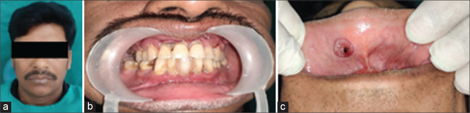
- (a) A 40-year-old male with a localized growth in the upper labial mucosa, (b) local foci of irritation, and (c) a solitary exophytic sessile growth was seen on upper labial mucosa.
On examining clinically, a solitary, exophytic, and sessile growth was appreciated on upper labial mucosa irt 11,12 measuring 1 cm approximately with pseudomembranous surface and area of erythema with smooth surface, regular border [Figure 1b and c].
On palpation, the mass was soft to firm in consistency and bleeded readily on provocation. Lymph nodes were non-palpable with no tenderness. Differential diagnosis of traumatic fibroma and peripheral giant cell granuloma was considered.
Pre-excision blood investigations were all within moderation except for the random blood sugar which was suggestive of pre-diabetic stage (94 mg/dL).
Oral prophylaxis was done to cut out local foci of irritation and infection. To minimize bleeding and reducing the chances of post-excision infection at the site, LASER of 940 nm was used.
The excised tissue was dispatched for histopathological examination which disclosed superficially ulcerated stratified squamous epithelium underlying fibrovascular and inflamed C.T. stroma which was loosely arranged and consisted of numerous proliferating endothelial lined blood vessels, spindle shaped fibroblasts, and chronic inflammatory infiltrates – lymphocytes and plasma cells and few PMNLs and extravasated RBCs. Pathologist impression was that of “PYOGENIC GRANULOMA.”
DISCUSSION
Occurrence of pyogenic granuloma was first brought in to light by Poncet and Dor in 1897 and was then named as botryomycosis hominis. The name “pyogenic granuloma” was first given by Hartzell in 1904. Over the span of time, this lesion has been suggested with various names as benign vascular tumor, Crocker and Hartzell’s disease, epulis teleangiectaticum granulomatosa, granuloma gravidarum or pregnancy tumor, hemangiomatosis granuloma, vascular epulis, and lobular capillary hemangioma.[1]
Among all the reactive lesions described, incidence rate of PG ranges between 26.8% and 32%. About 75% of PG cases in oral cavity are seen most commonly involving gingiva. In 5% of pregnancies, PG of the gingiva develops so terms like “pregnancy tumor” or “granuloma gravidarum” are often used. Extragingival sites are rarely involved.[2]
Pyogenic granuloma though effects all age groups has greater propensity in children and young adults.[3] Various contradicting reports exist regarding epidemiological pattern of disease. The vascular effects of female hormones justify a definite predilection which is demonstrated in most of the studies.[4]
Mechanism put forward emphasizes the significance of insults leading to disparity of anti-angiogenic factors and pro-angiogenic, causing a boom of neovascular capillaries which are friable and lobulated. History of trauma is directly attributed only in 7% lesions, reactive granulation from minor trauma may also be considered as contributory factor. Those consuming tacrolimus and cyclosporine after transplant of hematopoietic stem cell show up with intraoral pyogenic granuloma.[4]
The typical clinical presentation of pyogenic granuloma is small and vivid red to reddish-purple lesion seen on gingiva that is either pedunculated or sessile. The surface can be smooth, warty or lobulated, and it commonly gets ulcerated and prone to bleeding either spontaneously or as a result of slight minor trauma. Usually, the lesion is painless and soft in consistency and older lesions become more firm due to collagenization. The lesion is normally between 0.5 cm and 2 cm in size and can expand at an alarming rate, outstretching the size in a duration of week.[5] A lesion similar to clinical presentation of our case has been put up in literature by Kamala et al.[6]
Despite the fact that pyogenic granuloma is usually clinically diagnosed accurately, radiographic and histological examination helps in confirmation of diagnosis and guiding treatment plan.[7] To eliminate presence of bony destruction that could indicate malignancy or to spot a sharp restorative edge or foreign body that is supposed to be removed alongside lesion, radiographs are indicated. Dystrophic calcifications can also be seen in long-standing pyogenic granulomas. Radiographic appearance of these calcifications ranges from barely discernible fine radiopaque grains to larger and irregular radiopaque particles that rarely exceed 0.5 cm in diameter.[8]
Pyogenic granuloma’s microscopic picture in general reveals atrophic or hyperplastic epithelium covering exuberant granulation tissue, epithelium may ulcerate at times showing fibrinous exudates. The characteristic features of pyogenic granuloma are presence of voluminous endothelium-lined vascular spaces with fibroblastic proliferation and area of budding endothelial cells and infiltrating mixed inflammatory cells.[3] Cawson et al.[9] delineated pyogenic granuloma into two types based on vascularity and proliferation rate as: (i) Lobular capillary hemangioma and (ii) non-lobular capillary hemangioma.
In non-cosmetically sensitive areas, surgical excision and closure by suturing are considered mainstay for treatment in small lesion; whereas cauterization by silver nitrate should be considered treatment of choice under non-surgical modalities of treatment.[10]
Due to minimal side effects and rapid healing, Laser excision of PG is well warranted. Complete resolution of PG has been assisted by cryosurgery that shows good esthetic results. Furthermore, sclerosing agent (monoethanolamine oleate), ethanol, and multiple local intralesional steroid injection result in the complete resolution, without recurrence though multiple session is required for it.[11]
Even after surgical excision up to 16% of the lesions reoccurs.
CONCLUSION
Pyogenic granuloma can always occur on unexcepted site making diagnosis complex and also due to their clinical resemblances to other lesions of oral cavity, which entails a thorough histopathological examination of excised specimen to establish final diagnosis and institute an acceptable treatment.
Declaration of patient consent
The authors certify that they have obtained all appropriate patient consent.
Financial support and sponsorship
Nil.
Conflicts of interest
There are no conflicts of interest.
References
- Oral pyogenic granuloma: Various concepts of etiopathogenesis. J Oral Maxillofac Pathol. 2012;16:79-82.
- [CrossRef] [PubMed] [Google Scholar]
- Extragingival pyogenic granuloma of the lower lip masquerading as a vascular lesion. J Oral Maxillofac Pathol. 2022;26:119-23.
- [CrossRef] [PubMed] [Google Scholar]
- Oral and Maxillofacial Pathology (2nd ed). United States: WB. Saunders Co.; 2004. p. :444-9.
- [Google Scholar]
- Pyogenic granuloma In: StatPearls. Treasure Island, FL: StatPearls Publishing; 2022.
- [Google Scholar]
- Extragingival pyogenic granuloma. Indian J Dent Res. 2006;17:199-202.
- [CrossRef] [PubMed] [Google Scholar]
- Pyogenic granuloma on the upper labial mucosa: A case report. J Clin Diagn Res. 2013;7:1244-6.
- [Google Scholar]
- Extragingival pyogenic granuloma: An unusual clinical presentation. J Dent Shiraz Univ Med Sci. 2015;16:282-5.
- [Google Scholar]
- Oral Radiology Principles and Interpretation (5th ed). Missouri: Mosby; 2004. p. :598.
- [Google Scholar]
- Lucas Pathology of Tumors of Oral Tissues (5th ed). Missouri: Mosby; 1998. p. :252-4.
- [Google Scholar]
- Treatment options for cutaneous pyogenic granulomas: A review. J Plast Reconstr Aesthet Surg. 2011;64:1216-20.
- [CrossRef] [PubMed] [Google Scholar]
- Comparison of cryotherapy and curettage for the treatment of pyogenic granuloma: A randomized trial. Br J Dermatol. 2006;154:671-5.
- [CrossRef] [PubMed] [Google Scholar]


